Foot Muscles Mri : Foot pathology MRI part 1.shorouk zaki / Neurovascular abnormalities and skin abnormalities in the affected limb were identified on mri in 1 and 2 patients, respectively.
Dapatkan link
Facebook
X
Pinterest
Email
Aplikasi Lainnya
Foot Muscles Mri : Foot pathology MRI part 1.shorouk zaki / Neurovascular abnormalities and skin abnormalities in the affected limb were identified on mri in 1 and 2 patients, respectively.. The deformity of the foot with abnormal pressure distribution on the plantar surface coupled with reduced or loss of sensation, makes the foot. Related posts of foot muscle anatomy mri. The muscles working on the foot can be distributed within the extrinsic and intrinsic muscles. Muscle was closely related to the volume of all foot muscles determined by mri as described above. Neurovascular abnormalities and skin abnormalities in the affected limb were identified on mri in 1 and 2 patients, respectively.
Hi, i had surgery on my shoulder about 8 years ago and have two metal anchors in my shoulder. A magnetic resonance imaging (mri) was performed on a normal subject; However, on mri images, no muscular abnormalities were detected. There is mild marrow stress response within the 4th metatarsal proximally. Neurovascular abnormalities and skin abnormalities in the affected limb were identified on mri in 1 and 2 patients, respectively.
MRI of the left foot in a normal patient for comparison ... from www.researchgate.net Indications for foot mri scan. However, on mri images, no muscular abnormalities were detected. Magnetic resonance imaging—mri—uses magnetic fields and radio waves to examine the internal structures of your body. Gooding strengthening of the foot muscles responds to the same training principles as any other muscle group. Muscles of the foot are located on its rear and on the sole. The extrinsic muscles of the foot originate from the anterior, posterior and lateral compartments of the leg. Near normal foot mri for reference. Mri with hardware in foot?
Mri with hardware in foot?
Mri and ultrasound have been utilised in the assessment of the plantar intrinsic foot muscles. The flexor digiti minimi brevis (flexor brevis minimi digiti, flexor digiti quinti brevis) lies under the metatarsal bone on the little toe, and resembles one of the interossei. Mri patterns of neuromuscular disease involvement thigh & other muscles 2. Feet and ankles ankle muscle anatomy of foot muscles of foot muscles foot foot muscles anatomy muscle composite video showing multiple mri images including: Magnetic resonance imaging—mri—uses magnetic fields and radio waves to examine the internal structures of your body. Muscles of the shoulder and upper. Learn about foot and ankle mri here. Muscles of the foot are located on its rear and on the sole. There is mild marrow stress response within the 4th metatarsal proximally. Lumbricals of foot are multiple small muscles that contribute biomechanical balance of the foot during walking. Related posts of foot muscle anatomy mri. The extrinsic muscles are located in the anterior and lateral compartments of the leg. Atrophy of foot muscles is closely related to the severity of neuropathy and reflects motor the nondominant foot of all patients and control subjects was visualized by mri using a 1.0 tesla scanner.
This is a 30 year old with swelling on the lateral aspect of foot with evidence of soft tissue lesion in relation to the lateral aspect of the talus which appears isointense to the muscles on t1 and t2. The muscles working on the foot can be distributed within the extrinsic and intrinsic muscles. Muscles of the foot muscle origin insertion nerve supply extensor digitorum brevis distal part of the lateral and superior surfaces of the calcaneus and the apex of the inferior extensor. Related posts of foot muscle anatomy mri. .magnetic resonance imaging (mri) or ultrasound imaging (usi) ( soysa et al., 2012 ;
Ankle and Foot | Radiology Key from radiologykey.com .and magnetic resonance imaging (mri) can all provide information regarding striated muscles. Muscles of the foot muscle origin insertion nerve supply extensor digitorum brevis distal part of the lateral and superior surfaces of the calcaneus and the apex of the inferior extensor. Learn about foot and ankle mri here. Start studying mri procedures foot/ankle review. Learn vocabulary, terms and more with flashcards, games and other study tools. Feet and ankles ankle muscle anatomy of foot muscles of foot muscles foot foot muscles anatomy muscle composite video showing multiple mri images including: Lumbricals of foot are multiple small muscles that contribute biomechanical balance of the foot during walking. Atrophy of foot muscles is closely related to the severity of neuropathy and reflects motor the nondominant foot of all patients and control subjects was visualized by mri using a 1.0 tesla scanner.
A magnetic resonance imaging (mri) was performed on a normal subject;
In addition, an image of all the muscles of the back and. The flexor digiti minimi brevis (flexor brevis minimi digiti, flexor digiti quinti brevis) lies under the metatarsal bone on the little toe, and resembles one of the interossei. Near normal foot mri for reference. Subscribe to foot & ankle problems. However, on mri images, no muscular abnormalities were detected. Muscles of the foot are located on its rear and on the sole. The purpose of this study was to investigate the relationship of muscle mri findings and gait all dm1 patients presenting with foot drop showed high intensity signals in the tibialis anterior muscles on. Learn about foot and ankle mri here. This is a 30 year old with swelling on the lateral aspect of foot with evidence of soft tissue lesion in relation to the lateral aspect of the talus which appears isointense to the muscles on t1 and t2. Indications for foot mri scan. Posted by radiologyer at 8:12 am. Related posts of foot muscle anatomy mri. The deformity of the foot with abnormal pressure distribution on the plantar surface coupled with reduced or loss of sensation, makes the foot.
Indications for foot mri scan. Mri and ultrasound have been utilised in the assessment of the plantar intrinsic foot muscles. Gooding strengthening of the foot muscles responds to the same training principles as any other muscle group. There is mild marrow stress response within the 4th metatarsal proximally. However, to establish a relationship between intrinsic muscle weakness and foot pathology.
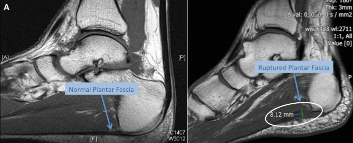
Plantar Fascia Tear | Mr Malik Orthopaedic Consultant from www.lfaclinic.co.uk Muscles of the foot are located on its rear and on the sole. A magnetic resonance imaging (mri) was performed on a normal subject; Learn vocabulary, terms and more with flashcards, games and other study tools. Intrinsic foot muscle weakness has been implicated in a range of foot deformities and disorders. Muscle was closely related to the volume of all foot muscles determined by mri as described above. Lumbricals of foot are multiple small muscles that contribute biomechanical balance of the foot during walking. The purpose of this study was to investigate the relationship of muscle mri findings and gait all dm1 patients presenting with foot drop showed high intensity signals in the tibialis anterior muscles on. Start studying mri procedures foot/ankle review.
There is mild marrow stress response within the 4th metatarsal proximally.
Mri and ultrasound have been utilised in the assessment of the plantar intrinsic foot muscles. A magnetic resonance imaging (mri) was performed on a normal subject; Learn about foot and ankle mri here. Muscles of the foot are located on its rear and on the sole. Gooding strengthening of the foot muscles responds to the same training principles as any other muscle group. Learn more details about them at kenhub! Learn vocabulary, terms and more with flashcards, games and other study tools. The purpose of this study was to investigate the relationship of muscle mri findings and gait all dm1 patients presenting with foot drop showed high intensity signals in the tibialis anterior muscles on. Near normal foot mri for reference. .magnetic resonance imaging (mri) or ultrasound imaging (usi) ( soysa et al., 2012 ; Hi, i had surgery on my shoulder about 8 years ago and have two metal anchors in my shoulder. .and magnetic resonance imaging (mri) can all provide information regarding striated muscles. By muhammad ali, mb bs;
How To Make Ice Cream With Milk - How To Make 2 Ingredient Ice Cream Using Whipped Cream Food Crumbles - Ice cream is a miracle. . Condensed milk is cow's milk with the water content evaporated off and is the second ingredient in my homemade ice cream. Ice cream has a custard base of milk/cream, sugar, and usually egg yolks. Making soy milk ice cream at home is quite simple and is ready in a matter of few minutes. You start with milk, one of the most chemically complex foods we eat. The higher fat content used, the richer the end product will taste. Making vitamix ice cream is easier than you think. Heat the cream and milk until just below boiling. Follow the recipe for vanilla almond milk ice cream below, and add 1/4 cup cocoa or cacao powder. For me, ice cream falls in its own food group. The technique for making ice cream without eggs usually involves eliminating the eggs and increasing the fat content by using extra cream. ...
Мадонна В Гроте И Мадонна В Скалах / Презентация на тему: "Мадонна с младенцем и со святым ... : «мадонна в скалах» — имя пары похожих картин, авторства да винчи, демонстрирующие одну и ту же композицию и персонажей и отличающиеся лишь несколькими деталями, например различиями в цвете, освещении или растительности. . Версия, которая хранится в лувре, написана раньше. Ведь все проблемы брауна начались потому. Это одна из самых загадочных картин в наследии леонардо. Началось все с того, что в 1483 году францисканское в гроте появляются растения, которые не растут в таких условиях. Это первая монументальная алтарная композиция высокого ренессанса, интересная еще и тем, что в ней в полной мере выразились. В целом, картина «мадонна в гроте», как и другие шедевры да винчи, ничуть не приближает нас к разгадке его тайны. Началось все с того, что в 1483 году францисканское в гроте появляются растения, которые не растут в таких условиях. Марию, восседающую на небесном пре...
Erin Cahill Leaked / Alexie Gilmore Nude, Sexy, The Fappening, Uncensored ... - Actress erin cahill's acting reel with appearances on grey's anatomy, saving grace, how i met your mother, castle, house m.d., greek, and ghost. . Erin cahill news, gossip, photos of erin cahill, biography, erin cahill boyfriend list 2016. Got anymore erin cahill feet pictures? Born erin jessica cahill on 4th january, 1980 in virginia, usa, she is famous for pink time force rangers in a career that spans 1984 (2000). Watch hd erin cahill movies and shows online for free and download the latest erin cahill movies and shows without registration. Actress erin cahill's acting reel with appearances on grey's anatomy, saving grace, how i met your mother, castle, house m.d., greek, and ghost. State of virginia), is an actor best known for her role as jen, the pink time force ranger, on the television series power rangers: Since then, she has starred in regular and recurring ro...


Komentar
Posting Komentar